Right Upper Back Anatomy : Upper Buttock Pain Sacro Illiac Joint Area Pain / It is very stiff, and the thoracic spine has a limited range of motion.
byAdmin-
0
Right Upper Back Anatomy : Upper Buttock Pain Sacro Illiac Joint Area Pain / It is very stiff, and the thoracic spine has a limited range of motion.. Away from, farther from the origin proximal: Sometimes you feel the effects right away. There is a set of muscles in the upper back (called the thoracic area) called the spinalis thoracis. The cervical spine supports the weight and movement of your head and protects the nerves exiting your brain. See back muscle anatomy stock video clips.
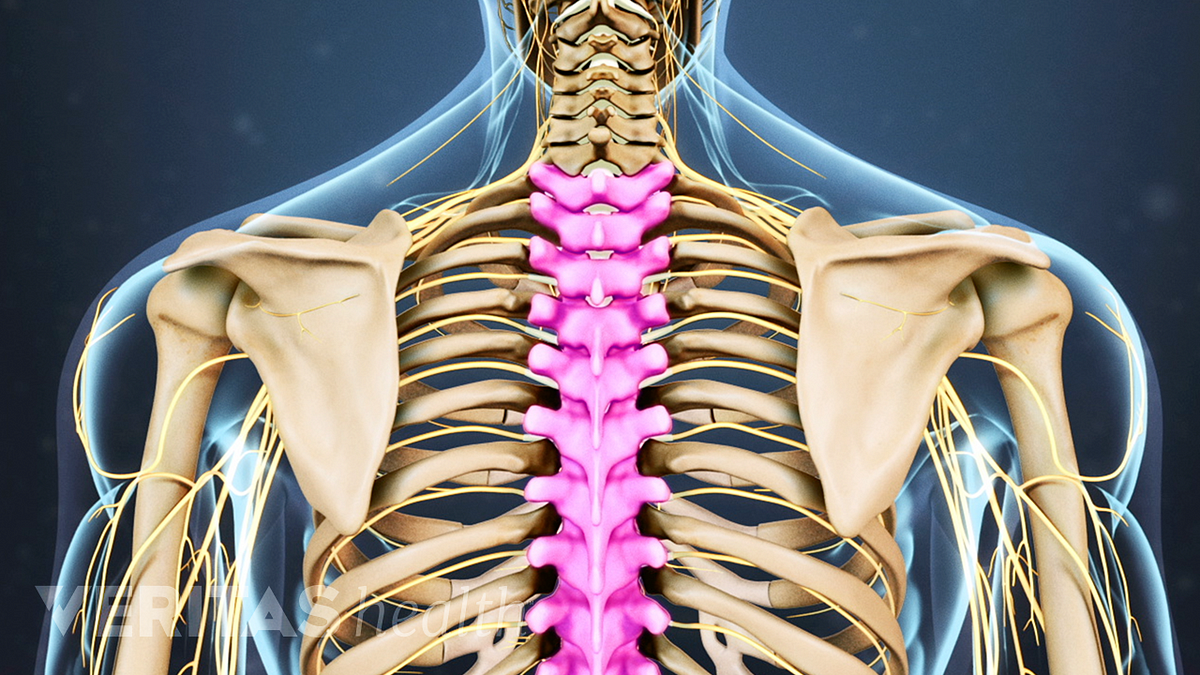
Balance the weight of your head on top of your spine. Connecting with the cervical spine above and the lumbar spine below, the thoracic spine runs from the base of the neck down to the abdomen. Human body anatomy female female anatomy muscle shoulder blade pain anatomy back muscles bones man female anatomy body muscles in a body female anatomy muscole shoulder concept muscular sysyem. It runs from the neck to the upper back. Human anatomy · july 23, 2016.
Thoracic Spine Anatomy And Upper Back Pain from embed.widencdn.net Symptoms of pulled upper back muscles may include: Limited range of motion or a gradual decrease in muscle movement. It is the only spinal region attached to the rib cage. These two terms, used in anatomy and embryology, describe something at the back (dorsal) or front/belly (ventral) of an organism. The anatomy of your back muscles can be complex. Thoracic spine anatomy and upper back pain. The cervical spine protects the nerves connecting to. This portion also attaches to the back part of the transverse processes of.
The iliocostalis muscles are furthest from the spine.
Your lower back (lumbar spine) is the anatomic region between your lowest rib and the upper part of the buttock. The cervical spine is the top part of the spine. 1 your spine in this region has a natural inward curve. This area of the body includes the top of the thoracic spine, which. Symptoms of pulled upper back muscles may include: In order to better visualize the common bile duct, the sonographer first identifies the portal vein in. Your back consists of a complex array of bones, discs, nerves, joints, and muscles. The thigh bears much of the load of the body's weight when a person is upright. It is the only spinal region attached to the rib cage. The cervical spine protects the nerves connecting to. It contains many muscles and nerves but only has one bone, the femur, which is the longest and strongest bone in. The cervical spine supports the weight and movement of your head and protects the nerves exiting your brain. It is like that for several reasons, all of which you can understand by looking at the anatomy of the thoracic spine.
Both the deltoid and the trapezius are firmly attached to … There is a set of muscles in the upper back (called the thoracic area) called the spinalis thoracis. Human body anatomy female female anatomy muscle shoulder blade pain anatomy back muscles bones man female anatomy body muscles in a body female anatomy muscole shoulder concept muscular sysyem. Sometimes you feel the effects right away. In order to better visualize the common bile duct, the sonographer first identifies the portal vein in.
Upper Back Pain Right Side Above Hip from pnpfitness.com Human musculature bodybuilding infographic muscular system vector human anatomy back muscle anatomy bicep male muscular anatomy human body anatomy female female anatomy muscle hamstrings muscle. It runs from the neck to the upper back. It contains many muscles and nerves but only has one bone, the femur, which is the longest and strongest bone in. The lumbar portion of the iliocostalis muscle travels upward from the lower area of the pelvis and sacrum to attach onto the lower border of the bottom six or seven ribs, by means of tendons that branch off from the main line.; Right upper back pain can be due to problems affecting the lungs, particularly if the right lung is involved. License image the deltoid, teres major, teres minor, infraspinatus, supraspinatus (not shown) and subscapularis muscles (not shown) all extend from the scapula to the humerus and act on the shoulder joint. Near, closer to the origin dorsal: After, behind, following, toward the rear distal:
The cervical spine protects the nerves connecting to.
The upper right quadrant of the abdomen contains the liver, the gallbladder, the duodenum, the head of the pancreas, the right kidney and the part of the small intestine that connects to the stomach (duodenum). If talking about the skull, the dorsal side is the top. The lung tissue of this lobe is responsible for most of the gas exchange in the right lung during calm, shallow breathing. Related posts of upper back muscle diagram muscle anatomy diagram. Your lower back (lumbar spine) is the anatomic region between your lowest rib and the upper part of the buttock. Back muscles anatomy here include the trapezius, latissimus dorsi, rhomboid and levator scapulae. The thigh bears much of the load of the body's weight when a person is upright. Human body anatomy female female anatomy muscle shoulder blade pain anatomy back muscles bones man female anatomy body muscles in a body female anatomy muscole shoulder concept muscular sysyem. Limited range of motion or a gradual decrease in muscle movement. It contains many muscles and nerves but only has one bone, the femur, which is the longest and strongest bone in. The lumbar and sacrum region make up the bone of the lower back anatomy. Doctors from medicinenet say that the middle and upper part of your spine contains 12 vertebrae that are attached to your rib cage. It runs from the neck to the upper back.
It comprises the vertebral column (spine) and two compartments of back muscles; The iliocostalis muscles are furthest from the spine. The back is the body region between the neck and the gluteal regions. See human back anatomy stock video clips. Away from, farther from the origin proximal:
Back Muscles Anatomy Of Upper Middle Lower Back Pain In Diagrams Goodpath from images.ctfassets.net Human musculature bodybuilding infographic muscular system vector human anatomy back muscle anatomy bicep male muscular anatomy human body anatomy female female anatomy muscle hamstrings muscle. These sections are cervical (neck), thoracic (upper and middle back), lumbar (lower back), and sacrum (tailbone). Related posts of upper back muscle diagram muscle anatomy diagram. Your back consists of a complex array of bones, discs, nerves, joints, and muscles. The back is the body region between the neck and the gluteal regions. These important muscles control many motions that involve moving the arms and head — such as throwing a ball, looking up at the sky, and raising your hand. See back muscle anatomy stock video clips. The liver is the largest abdominal organ responsible for multiple metabolic processes of the body.
The thoracic spine is the longest region of the spine, and by some measures it is also the most complex.
The hurt can stem from sore muscles, ligaments, and tendons, or from herniated disks, fractures, and other problems in your upper, middle, and lower back. The upper arm is located between the shoulder joint and elbow joint. The thoracic portion also attaches to ribs, but these are the top part of the upper six ribs. In order to better visualize the common bile duct, the sonographer first identifies the portal vein in. The lung tissue of this lobe is responsible for most of the gas exchange in the right lung during calm, shallow breathing. The liver is the largest abdominal organ responsible for multiple metabolic processes of the body. Common causes include pneumonia and pleurisy, which refers to infection or inflammation of the thin layer of tissue that surrounds the lung and lines the chest cavity. The human spine is composed of 4 sections of vertebrae. Doctors from medicinenet say that the middle and upper part of your spine contains 12 vertebrae that are attached to your rib cage. License image the deltoid, teres major, teres minor, infraspinatus, supraspinatus (not shown) and subscapularis muscles (not shown) all extend from the scapula to the humerus and act on the shoulder joint. Thoracic spine anatomy and upper back pain. It runs from the neck to the upper back. The back functions are many, such as to house and protect the spinal cord, hold the body and head upright, and adjust the movements of the upper and lower limbs.